Seaweed disease: Ice-ice On Seaweed
Cause: Environmental factors and several types of bacteria: Pseudoalteromonas gracilis, Pseudomonas spp .. and Vibrio spp.
Bio - Ecology Pathogens:
• The case of ice-ice on seaweed farming is triggered by fluctuations in water quality parameters are extreme (salinity, water temperature, dissolved organic matter and sunlight intensity).
• Other triggers are insect like fish baronang, green turtles, sea urchins and starfish cause injury to the thallus, so easily infected by microorganisms.
• In the state of stress, sea grass will release organic substances that cause thallus slimy and stimulate the bacteria grow abundantly in the vicinity.
• Growth of bacteria on the thallus will cause the thallus becomes white and fragile. Furthermore, easily broken, and the soft tissue that characterize the ice-ice disease.
• The spread of this disease can occur vertically (from seed) or horizontally through the mediation of water.
Clinical symptoms:
• The disease is characterized by the emergence of spots / red spots on some of the old thallus gradually became pale yellow and finally fade to white. Thallus become brittle and easily broken.
• Symptoms are shown slow growth. the color change becomes pale and at several branches and rotting thallus to be white.
Diagnosis:
• visual and microbiological observations.
Control:
• The use of quality seeds is a very important way to control ice-ice disease.
• Disinfection of seedlings can be dipped in the solution by PK (potassium permanganate) with a dose of 20 PPM
• Selection of farms that meet the optimum requirements for growth of seaweed.
• Application of cultivation techniques adapted to aquatic environments
• Noting the season in relation to cultivation techniques that would be applied.
source: Ministry of Maritime Affairs and Fisheries of Indonesia, Directorate General of Aquaculture, Fish and Environmental Health Directorate, 2010
Tampilkan postingan dengan label fish disease. Tampilkan semua postingan
Tampilkan postingan dengan label fish disease. Tampilkan semua postingan
Selasa, 07 Juni 2011
Minggu, 05 Juni 2011
Fish Disease: vibriosis in shrimp
Fish Disease: vibriosis in shrimp
Cause: Vibrio harveyii, V. alginolyticus, V. parahaemolyticus. etc..
Bio - Ecology Pathogens
• vibriosis in shrimp larvae commonly as secondary infection, especially when under stress and weak.
• Bacterial infections are usually associated with stress conditions due to: high density, malnutrition, poor handling. parasitic infections, high organic matter, low oxygen. poor water quality. extreme fluctuations in water temperature. etc..
• The attack is acute, and if environmental conditions continue to decline, which caused the death can reach 100%. particularly in post-larvae or juvenile stage.
Clinical symptoms:
• Body of shrimp look dull and dirty.
• decreased appetite, damage to the legs and gills, gill or brownish color.
• Types of Vibrio spp. which generally attacks the larvae glow shrimp and prawn disease disease called glow (luminescent vibriosis).
• Shrimp affected showed symptoms of necrosis, the condition of the body is weak, slow swim, appetite loss, red spots (red discoloration) on the pleopod and abdominal as well as visible light at night
• Shrimp vibriosis affected leg will show the pool (pleopoda) and the foot path (pereiopoda) shows melanisasi.
• Shrimp are dying often swim to the surface or edge of the pond embankment.
Diagnosis:
• Isolation and identification of bacteria through bio-chemical tests.
Control:
• Disinfection of aquaculture facilities before and during the maintenance process shrimp
• Giving immunostimulan element (eg addition of
vitamin C in feed) are routinely during maintenance
• Avoiding the occurrence of stress (physical, chemical, biological)
• shrimp health management in an integrated manner
source: Ministry of Maritime Affairs and Fisheries of Indonesia, Directorate General of Aquaculture, Fish and Environmental Health Directorate, 2010
Minggu, 29 Mei 2011
fish disease: vibriosis in fish
fish disease: vibriosis in fish
Cause: Vibrio alginolyticus, V. parahaemolyticus, V. vulnificus, V. ordalii, etc..
Bio - Ecology Pathogens
• Bacteria in sea water ecosystems, and vibirosis still
is a major problem for marine fish farming industry.
• The case of vibriosis can occur throughout the year, but commonly associated with stress due to handling, high density or changes in extreme weather.
• The death rate of fish at the larval stage up to the size
fingerlings are attacked by the bacteria may reach 80-90%.
Clinical Symptoms:
• Weak, loss of appetite, swim in the water surface, and opaque color.
• Inflammation of the rectum, gills, mouth, base of fin, followed by bleeding and blisters on the surface of the body, as well as open wounds.
• In advanced infection of bleeding in the mouth and base of fins, excess mucus in the gills, dropsy, pale liver color. and eyes swollen.
Diagnosis
• Isolation and identification of bacteria through a bio-chemical tests
Control:
• Disinfection of aquaculture facilities before and during the maintenance of fish
• Giving immunostimulan element (eg addition of
vitamin C in feed) are routinely during maintenance
• Avoiding the occurrence of stress (physical, chemical, biological)
• Management of fish health in an integrated (fish, environment and pathogens)
• Limit and / or regulate feeding and mixing of feed with drugs (medicated feed and feed restriction)
• Conducting anti-vibriosis vaccination.
source: Ministry of Maritime Affairs and Fisheries of Indonesia, Directorate General of Aquaculture, Fish and Environmental Health Directorate, 2010
Minggu, 15 Mei 2011
fish disease: Enteric Septicemia of Catfish (ESC)
fish disease: Enteric Septicemia of Catfish (ESC)
Cause: Edwarsiella ictaluri
Bio-Ecology Pathogens:
• rod-shaped bacteria, gram-negative character moves with the aid of flagella. does not form spores or capsules and are facultative anaerobes.
• Originally known to only infect fish cannel catfish, but the latter is known to infect other fish species such as catfish, catfish, and eel. Experimentally, several types of fish such as trout, tilapia, salmon and ornamental fish can also be infected with this bacteria.
• Transmission of horizontally ie contact between the host or through the water.
• Case ESC generally occurs when water temperatures are relatively warm (22-28 degrees Celsius), but at water temperatures below 20 degrees Celsius or above 30 degrees centigrade, this bacteria is decreased malignancy.
Clinical Symptoms
• Weak, loss of appetite. color pale gills, sometimes protruding eyes and / or abdominal swelling (dropsy)
• Often, too, found the existence of petechiae (red spots) on the body part that is not pigmented (under the chin, stomach or at the base of the fin)
• Swim in the water or the pool with his head pointing up
• Before dying, the fish usually swim like a spastic and / or swim like a spiral spin
• There are white patches on internal organs (liver, spleen, kidney, d1l.)
Diagnosis
• Isolation and identification of bacteria through bio-chemical tests.
• Detection of bacterial genes by polymerase chain reaction (PCR)
Control:
• Avoiding the occurrence of stress (physical, chemical, biological)
• Improve overall water quality, particularly reducing the levels of dissolved organic material and / or increase the frequency of replacement of new water
• Management of fish health in an integrated (fish, environment and pathogens)
• Limit and / or regulate feeding and mixing of feed with drugs (medicated feed and feed restriction)
• Conducting anti-Edwardsiella ictaluri vaccine.
source: Ministry of Maritime Affairs and Fisheries of Indonesia, Directorate General of Aquaculture, Fish and Environmental Health Directorate, 2010
Selasa, 10 Mei 2011
Fish Disease: Edwarsiellosis
Fish Disease: Edwarsiellosis
Cause: Edwarsiella tarda
Bio-Ecology Pathogens:
• curved rod-shaped bacteria, gram-negative character moves with the aid of flagella, do not form spores or capsules, are facultative anaerobes, and able to produce H2S.
• Found in freshwater environments and sea water, infecting several species of fish include: salmon, catfish, carp, tilapia. etc..
• Transmission of horizontally ie contact between host one with another host or through water.
• Generally occurs at a relatively high water temperature (± 30 degrees Celsius) and high organic matter content.
• The death rate depends on environmental conditions, in very poor conditions can lead to death by 50%.
Clinical Symptoms:
• In mild infections, revealing only minor injuries.
• As the development of more advanced disease, purulent wound developed in the ribs and stomach muscles.
• Pale, bloated stomach containing a yellowish liquid or redness, bleeding of the rectum and / or depressed into the anus, and eyes faded.
• Further development, injuries (cavities) experience swelling and if scratched will smell of H2S gas.
Diagnosis:
• Isolation and identification of bacteria through bio-chemical tests.
• Detection of bacterial genes by polymerase chain reaction (PCR)
Control:
• Avoiding the occurrence of stress (physical, chemical, biological)
• Improve overall water quality, particularly reducing the levels of dissolved organic material and / or increase the frequency of replacement of new water
• Management of fish health in an integrated (fish, environment and pathogens)
• Limit and / or regulate feeding and mixing of feed with drugs (medicated feed and feed restriction)
• Conducting anti-Edwardsiella tarda vaccination.
source: Ministry of Maritime Affairs and Fisheries of Indonesia, Directorate General of Aquaculture, Fish and Environmental Health Directorate, 2010
Jumat, 08 April 2011
Bacterial Fin / Tail Rot / Pseudomoniasis
Bacterial Fin / Tail Rot / Pseudomoniasis
Cause: Pseudomonas spp.
BioEkologi Pathogens:
• It is a gram-negative bacteria and non-spore. These bacteria are aerobic. with a size of 3 um x 0.5 um, motile, producing fluorescent pigment. and breed in soil and water.
• Hazardous mainly on freshwater fish (although it also can attack sea fish and brackish) and can result in high mortality due to infectious disease in quick time when water conditions allow.
• Transmission and spread of disease through direct contact with fish that are sick or with the polluted environment.
• The attacks can occur when fish are vulnerable or weakened by hunger. the feed is not suitable. cold, or water conditions are not good.
Clinical Symptoms
• Fish weak to move slowly. breathe gasping at the surface of the water.
• Color pale gills and a dark body color change.
• There are patches of red on the outside of his body and damage to the fins, gills and skin
• excessive mucus at first, then emerged bleeding
• fin and tail loss (decayed)
• bleeding. stomach became bloated fish, known as dropsy.
Diagnosis:
• isolation and identification of bacteria through bio-chemical tests.
Control:
• Avoiding the occurrence of stress (physical, chemical, biological)
• Improve overall water quality, particularly reducing the levels of dissolved organic material and / or increase the frequency of replacement of new water
• Management of fish health in an integrated (fish, environment and pathogens)
• Reduce feeding and number of fish in pond
• Soaking in a solution of PK 20 ppm for 30 minutes.
source: Ministry of Maritime Affairs and Fisheries of Indonesia, Directorate General of Aquaculture, Fish and Environmental Health Directorate, 2010
Minggu, 20 Maret 2011
Disease Mycobacteriosis / Fish Tuberculosis (TB)
Disease Mycobacteriosis / Fish Tuberculosis (TB)
Cause: Mycobacterium marinum (sea water) and M. fortuitum (fresh water)
Bio - Ecology pathogen
• bacteria are gram positive, short rod-shaped and non-motile.
• rain-fed pool and garden with limited water resources are more susceptible to infection type of the disease.
• Shows symptoms varied, but often show no clinical symptoms at all.
• The pattern of attacks are chronic mycobacteriosis - sub acute, both in freshwater fish, brackish and sea water fish.
• The optimum temperature ranges from 25-35 ° C, but still can grow well at 18-20 ° C.
Clinical Symptoms:
• Loss of appetite, weak, thin, bulging eyes (exopthalmia) and swelling of the body.
• If the infected skin, red patches occur and develop into sores, fin and tail damage.
• In the advanced phase of infection, internally there has been swelling bile, kidneys and liver, and is often found in the tubercle / brownish white nodule.
• slow growth, pale color and not beautiful, especially for ornamental fish.
• Lordosis, scoliosis, ulcer and fin damage (fractures) can occur in some fish that was attacked.
Diagnosis:
• Isolation using selective media, and
identified through bio-chemical tests.
• Detection of bacterial genes by polymerase chain reaction (PCR)
Control:
• infected fish were taken and destroyed immediately
• Avoid using water from ponds that are infected with the bacteria.
• Improve overall water quality, particularly reducing the levels of dissolved organic material and / or - to increase the frequency of replacement of new water
• Management of fish health in an integrated (fish, environment and pathogens)
• Soaking chloramine B or T 10 ppm for 24 hours and after that turn of the new water.
source: Ministry of Maritime Affairs and Fisheries of Indonesia, Directorate General of Aquaculture, Fish and Environmental Health Directorate, 2010
Kamis, 17 Maret 2011
Streptococciasis Disease
Streptococciasis Disease
Cause: Streptococcus agalactiae, S. iniae,
Bio - Ecology of pathogens:
• gram-positive bacteria, small round (cocci), joined
chain-like, non-motile, transparent and smooth colonies.
• Streptococcus iniae Infection often occurs in sea water fish farming (snapper, grouper), whereas S. agalactiae is more commonly found in freshwater fish farming (tilapia).
• The pattern of attacks are generally two types of bacteria are chronic - acute.
• Target organs of infection of Streptococcus spp. commonly found in the brain and eyes. so-called "syndrome, meningoencephalitis and panophthalmitis". The disease is frequently reported in intensive aquaculture systems, aquatic environment calm (stagnant) and / or recirculation systems,
• Cumulatively, the attack of this disease can cause mortality of 30-100% of the total population during the maintenance period: and this disease is a potential obstacle that must be anticipated with respect to intensification and improvement of national tilapia production.
Clinical Symptoms:
• Indicates abnormal behavior such as convulsions or spinning and prominent eyes (exopthalmus).
• decreased appetite, weakness, dark-colored body, and slow growth.
• Dark colors under the jaw, prominent eyes, bleeding, abdominal bloat (dropsy) or injuries that develop into ulcers.
• Occasionally. do not show obvious clinical symptoms except death continues.
• no directional movement (nervous) and bleeding on the gill cover (operculum).
• Often, too, found that the infected fish appear normal until shortly before death.
Diagnosis
• Isolation and identification of bacteria through bio-chemical tests.
• Detection of bacterial genes by polymerase chain reaction (PCR)
Control:
• Disinfection of aquaculture facilities before and during the maintenance of fish
• Prevention of early (seed) through vaccination anti-Streptococcus spp.
• Giving immunostimulan element (eg addition of
vitamin C in feed) are routinely during maintenance
• Improve overall water quality, particularly reducing the levels of dissolved organic material and / or increase the frequency of replacement of new water
• Management of fish health in an integrated (fish, environment and pathogens)
source: Ministry of Maritime Affairs and Fisheries of Indonesia, Directorate General of Aquaculture, Fish and Environmental Health Directorate, 2010
Rabu, 16 Februari 2011
DISEASE BACTERIA (bacterial DISEASE)
DISEASE BACTERIA (bacterial DISEASE)
1. Red disease (Motile Aeromonas Septicemia)
Cause: Aeromonas hydrophila
Bio-Ecology Pathogens:
• It is a bacterial disease that often occurs at all ages and types of freshwater fish, although the type of bacteria often found in brackish water and marine fish.
• Bacterial infections are usually associated with stress conditions due to: high density, malnutrition, poor handling, poor water quality, extreme fluctuations in water temperature, etc..
• The attack is acute, and if environmental conditions continue to decline, which caused the death can reach 100%.
Clinical Symptoms
• The color of the body dull / dark, decreased appetite, gathered near the drain, visible skin, and excess mucus.
• Bleeding at the base of the fins, tail, around the anus and other body parts.
• Scales loose, sores around the mouth, and other body parts.
• In severe infections, flabby stomach and swelling (dropsy) containing a yellowish red liquid.
• suffocate fish commonly found in surface and bottom of the pond.
Diagnosis:
• Isolation and identification of bacteria through bio-chemical tests.
• Detection of bacterial genes by polymerase chain reaction (PCR)
Control:
• Prevention of early (seed) through vaccination anti Aeromonas hydrophila (HydroVac)
• Disinfection of aquaculture facilities before and during the maintenance of fish
• Giving immunostimulan element (eg addition of
vitamin C in feed) are routinely during maintenance
• Avoiding the occurrence of stress (physical chemistry. Biologist)
• Improve overall water quality, particularly reducing the levels of dissolved organic material and / or increase the frequency of replacement of new water
• Management of fish health in an integrated (fish, environment and pathogens)
• Oxolinic acid at a dose of 10 mg / kg body weight of fish / day for 10 days
source: Ministry of Maritime Affairs and Fisheries of Indonesia, Directorate General of Aquaculture, Fish and Environmental Health Directorate, 2010
Selasa, 25 Januari 2011
Lerniasis (fish disease)
Lerniasis (fish disease)
Cause: Lemaeae cyprinaceae and L. arcuata
Bio - Ecology:
• The parasite is known as anchor worm (anchor worm).
• Sticking to the body of the fish with the "anchor" the stabbing and develop under the skin.
• The parasite is equipped with two egg sacs will be seen hanging outside the body of the fish.
• Almost all freshwater fish species susceptible to this parasitic infection, particularly the size of the seed.
• At high levels of infection can lead to serious cases of death.

Clinical Symptoms:
• Looks like an arrow that pierced the body of the fish. Sometimes the moss parasites on the body so that the infected fish looks like carrying a green flag
• There was a wound or bleeding at the site where the patch. In fish seed puncture it can reach internal organs so that it can lead to death
Diagnosis:
• Visually looks a parasite that attach to the fish body
Control:
• Precipitation and filtering incoming water.
• Destruction of infected fish and draining the pond, followed by calcification.
• Soaking with:
✓ formalin solution at 250 ppm for 15 minutes.
✓ Abate solution at a dose of 1 ppm (aquarium) and 1.5 ppm (Pool)
✓ dichlorvos solution 0.2 mg / L for 24 hours or more, every week for 4 consecutive weeks
sumber : Kementerian Kelautan dan Perikanan, Direktorat Jenderal Perikanan Budidaya, Direktorat Kesehatan Ikan dan Lingkungan, 2010
Cause: Lemaeae cyprinaceae and L. arcuata
Bio - Ecology:
• The parasite is known as anchor worm (anchor worm).
• Sticking to the body of the fish with the "anchor" the stabbing and develop under the skin.
• The parasite is equipped with two egg sacs will be seen hanging outside the body of the fish.
• Almost all freshwater fish species susceptible to this parasitic infection, particularly the size of the seed.
• At high levels of infection can lead to serious cases of death.

Clinical Symptoms:
• Looks like an arrow that pierced the body of the fish. Sometimes the moss parasites on the body so that the infected fish looks like carrying a green flag
• There was a wound or bleeding at the site where the patch. In fish seed puncture it can reach internal organs so that it can lead to death
Diagnosis:
• Visually looks a parasite that attach to the fish body
Control:

• Precipitation and filtering incoming water.
• Destruction of infected fish and draining the pond, followed by calcification.
• Soaking with:
✓ formalin solution at 250 ppm for 15 minutes.
✓ Abate solution at a dose of 1 ppm (aquarium) and 1.5 ppm (Pool)
✓ dichlorvos solution 0.2 mg / L for 24 hours or more, every week for 4 consecutive weeks
sumber : Kementerian Kelautan dan Perikanan, Direktorat Jenderal Perikanan Budidaya, Direktorat Kesehatan Ikan dan Lingkungan, 2010
Jumat, 21 Januari 2011
Benediasis
Benediasis
Cause: Benedinia sp. and Neo Benedinia sp.
Bio-Ecology Pathogens:
• blood eaters' blood feeder ", infect marine fish, especially snapper, and grouper.
• Parasites belonging to the Capsalid monogenea, which is a kind of worm trematoda.
• serious cases generally occur in fish culture in floating net (KJA).
• If the eye can cause blindness, and the resulting wound is the entrance for secondary bacterial infections.
• Deaths due to heavy infection can reach 30%.
Clinical symptoms:
• Wounds and bleeding at the site of the bite, and this looks visually parasites attached to the body of the fish, especially on the scales or on the fin (visible after the infected fish marinated in fresh water for several minutes)
• In severe infection the parasite can infect the eye, so that the eyelets will look white.
Diagnosis:
• Visually looks a parasite that attach to the fish's body, if placed in freshwater fish
Control:
• shed the parasite in the container is limited by using fresh water for 2-5 minutes.
• Soaking in a solution of hydrogen peroxide (H2O2) at a dose of 150 ppm for 10-30 minutes.
• After the parasitic loss, the fish was transferred to another container to prevent any secondary infection by bacteria in the parasite bites.
source : Kementerian Kelautan dan Perikanan, Direktorat Jenderal Perikanan Budidaya, Direktorat Kesehatan Ikan dan Lingkungan, 2010
Cause: Benedinia sp. and Neo Benedinia sp.
Bio-Ecology Pathogens:
• blood eaters' blood feeder ", infect marine fish, especially snapper, and grouper.
• Parasites belonging to the Capsalid monogenea, which is a kind of worm trematoda.
• serious cases generally occur in fish culture in floating net (KJA).
• If the eye can cause blindness, and the resulting wound is the entrance for secondary bacterial infections.
• Deaths due to heavy infection can reach 30%.
Clinical symptoms:
• Wounds and bleeding at the site of the bite, and this looks visually parasites attached to the body of the fish, especially on the scales or on the fin (visible after the infected fish marinated in fresh
 water for several minutes)
• In severe infection the parasite can infect the eye, so that the eyelets will look white.
Diagnosis:
• Visually looks a parasite that attach to the fish's body, if placed in freshwater fish
Control:
• shed the parasite in the container is limited by using fresh water for 2-5 minutes.
• Soaking in a solution of hydrogen peroxide (H2O2) at a dose of 150 ppm for 10-30 minutes.
• After the parasitic loss, the fish was transferred to another container to prevent any secondary infection by bacteria in the parasite bites.
source : Kementerian Kelautan dan Perikanan, Direktorat Jenderal Perikanan Budidaya, Direktorat Kesehatan Ikan dan Lingkungan, 2010
Rabu, 22 Desember 2010
Dactylogyriasis (Worms Gills)
Dactylogyriasis (Worms Gills)
Cause: Dactylogyrus spp., Cychlidogyrus spp., Quadricanthus spp.
Bio-Ecology Pathogens

• Ekto-obligate parasites that are parasitic and reproduce by laying eggs
• infect all species of freshwater fish, especially the size of the seed. Transmission occurs when infective face (Onchomiracidium).
• Dactylogyrus spp. has 2 pairs of eye point, and at the tip of his head there are 4 bumps. Cychlidogyrus spp. shape is more flattened at both ends, and only has a pair of eye point. Quadricanthus spp. shape
Dactylogyrus similar spp., and has a host of species that target specific groups of catfish.
• Severe infections can kill 30-100% within a few weeks
Clinical Symptoms:
• pale body color, decreased appetite, thin, nervous and slow
• Respiratory frequency increased, the production of excess mucus in the gills and often cavort
• Gather / closer to the water inlet
• Gills pale or swollen so that the open operculum
Diagnosis:
• Visual observation of behavior and clinical symptoms that arise
• Microscopic observation to see morphology
parasites through the production segment of the organ gill preparations.
Control:
• Maintaining water quality, especially the stabilization of the water temperature> 29 degrees Celsius
• Reducing the levels of dissolved organic material and / or increase the frequency of water changes
• Dactylogyriasis attacked fish with prevalence and intensity level is low, treatment can be done by soaking several types of disinfectants, among others:
✓ salt solution at a concentration 500-10000
ppm (depending on the type and age of fish) for 24 hours
✓ Solution Potassium Permanganate (PK) at a dose of 4 ppm for 12 hours
✓ formalin solution at doses of 25-50 ppm for 24 hours or more
✓ Glacial acetic acid 0.5 ml / L for 30 seconds every 2 days for 3 - 4 times
source: Ministry of Maritime Affairs and Fisheries of Indonesia, Directorate General of Aquaculture, Fish and Environmental Health Directorate, 2010
Kamis, 25 November 2010
Microsporidiasis (Cotton Shrimp Disease)
Microsporidiasis (Cotton Shrimp Disease)
Cause: The Microsporidia of the genera Thelohania, Nosema and Peistophora

Bio - Ecology Pathogens
• Named as cotton shrimp disease and / or shrimp milk.
• Having more than 8 spores in each capsule
• Virtually all penaeid shrimp species was reported the least susceptible to infection one type of parasite microsporidia group, although there are indications of specific local
• low pathogenicity, prevalence rates in a population generally not more than 5% and the resulting mortality was also relatively low
Clinical symptoms:
• Parts of the body of infected shrimp white milk and more soft
• white spores spread on the meat / muscle (internal parasites)
• Shrimp weak, easy to stress, decreased appetite, making it easy prey to predators sluggish, and easily die after handling (handling)

Diagnosis:
• Visual observation of behavior and clinical symptoms are quite clear
• Microscopic observation to see the morphology of microsporidia by making preparations for review of target organ infection. The observation that more clear on the characteristics of spores required specific staining.
Control:
• disinfection, drying of pond bottom and water sources that are free of microsporidia
• Shrimp are infected immediately destroyed, in order to reduce the potential for horizontal transmission
• To cut the parasite's life cycle, avoiding the feeding of trash fish infected with microsporidia
• No chemicals are effective for preventing and / or treat diseases microsporidiasis.
source : Kementerian Kelautan dan Perikanan, Dirjen. Perikanan Budidaya, 2010
Selasa, 16 November 2010
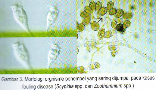
Filthy disease (fouling Disease)
Filthy disease (fouling Disease)
Cause: Zoothamnium spp., Epistylis spp., Vorticella spp.,. Acineta spp.
Bio - Ecology Pathogens
• Generally caused by microorganisms of the group
Protozoa, although often associated with algae
such as Nitzschia spp., Amphiprora spp., Navicula spp.,
Enteromorpha spp., Etc.
• Complex infection will interfere with the movement of microorganisms, especially shrimp larvae, difficulty eating, swimming, and the process of molting because gill organ and / or whole body filled with penempel organisms.
• Factors triggering the explosion of the disease, among others, high density, malnutrition, high levels of organic matter, and fluctuations in water quality parameters, especially temperature
Clinical Symptoms:
• Swim to the surface water and body-colored opaque / dirty
• Gills are infected with reddish or brownish color
• Weak, difficulty breathing and decreased appetite, finally dies
• The process of molting (moulting) are blocked, and the resulting inflammation of the skin
Diagnosis:
• Visual observation of behavior and clinical symptoms that arise
• Microscopic observation to see the morphology of organisms through the production of preparations penempel review of organ skin / mucus, fins and / or gills.
Control:
• Disinfection of containers / plot maintenance and sources of water that is free of microorganisms penempel)
• Improve overall water quality, particularly reducing the levels of dissolved organic material and / or increase the frequency of replacement of new water
• Giving immunostimulan element (eg addition of
vitamin C in feed) are routinely during maintenance
• Stimulate the process of molting through manipulation of water quality parameters which is a determinant factor

• Shrimp are attacked by "fouling disease" by the level of
low prevalence and intensity, treatment can
performed with several types of disinfectants, among others:
✓ Soaking in a solution of formalin at doses of 25-50
ppm for 24 hours or more
source: Ministry of Maritime Affairs and Fisheries Republic of Indonesia, Director General. Aquaculture, 2010
Cause: Zoothamnium spp., Epistylis spp., Vorticella spp.,. Acineta spp.
Bio - Ecology Pathogens

• Generally caused by microorganisms of the group
Protozoa, although often associated with algae
such as Nitzschia spp., Amphiprora spp., Navicula spp.,
Enteromorpha spp., Etc.
• Complex infection will interfere with the movement of microorganisms, especially shrimp larvae, difficulty eating, swimming, and the process of molting because gill organ and / or whole body filled with penempel organisms.
• Factors triggering the explosion of the disease, among others, high density, malnutrition, high levels of organic matter, and fluctuations in water quality parameters, especially temperature
Clinical Symptoms:
• Swim to the surface water and body-colored opaque / dirty
• Gills are infected with reddish or brownish color
• Weak, difficulty breathing and decreased appetite, finally dies
• The process of molting (moulting) are blocked, and the resulting inflammation of the skin
Diagnosis:

• Visual observation of behavior and clinical symptoms that arise
• Microscopic observation to see the morphology of organisms through the production of preparations penempel review of organ skin / mucus, fins and / or gills.
Control:
• Disinfection of containers / plot maintenance and sources of water that is free of microorganisms penempel)
• Improve overall water quality, particularly reducing the levels of dissolved organic material and / or increase the frequency of replacement of new water
• Giving immunostimulan element (eg addition of
vitamin C in feed) are routinely during maintenance
• Stimulate the process of molting through manipulation of water quality parameters which is a determinant factor
• Shrimp are attacked by "fouling disease" by the level of
low prevalence and intensity, treatment can
performed with several types of disinfectants, among others:
✓ Soaking in a solution of formalin at doses of 25-50
ppm for 24 hours or more
source: Ministry of Maritime Affairs and Fisheries Republic of Indonesia, Director General. Aquaculture, 2010
Sabtu, 13 November 2010
Fish disease: Cryptocaryasis (Marine White Spot)
Cryptocaryasis (Marine White Spot)
Cause: Cryptocaryon irritans
Bio - Ecology phatogen:
• Shaped round or oval measuring between 0.3-0.5 mm, and have cilia.
• obligate parasitic nature (a character similar biology with parasites "Ich")
• Highly malignant, in heavy infection can kill up to 100% in a few days
• infect marine aquaculture fish species (grouper, snapper, baronang, d1l.) Particularly seed size, although the size of adults are also vulnerable when their immune decline

Clinical Symptoms:
• decreased appetite, thin, dark body color, restlessness, lethargy and weakness
• Rubbing the body-rub on a nearby object
• Respiratory frequency increased (gasp), get closer to the water in.
• white spots or brown in fins, skin or gills, excess mucus production, and fin furl
• In severe infection, white spots or looks like a snow accompanied by bleeding, and opaque eyes, causing blindness
• secondary infection by bacteria will exacerbate health conditions to accelerate the process of death.
Diagnosis:
• Observations are visually for the presence of white spots (parasite) on the skin, fins and gills of fish
• Microscopic observation to see the morphology of the parasite through the production segment of the organ preparations skin / mucus, fins and / or gills.
Control:
• Maintain the temperature for always> 29 degrees Celsius
• The transfer of parasite-infected fish population to the water
parasite-free as much as 2-3 times with intervals of 2-3 days.
• Treatment and / or eradication of parasites may
conducted using immersion:

✓ low salinity water (0-8 PROMIL) for several hours (depending on species and size), transferred to water that is free of parasites and repeated every 2-3 days
✓ solution of hydrogen peroxide (H2O2) at a dose of 150 ppm for 30 minutes, transferred to water that is free of parasites and repeated every 2 days
✓ solution of copper sulphate (CuSO4) at doses of 0.5 ppm for 5-7 days with strong aeration, and water must be replaced every day.
✓ 25-50 ppm formalin solution for 12-24 hours, made repeated every 2 days
sumber : Kementerian Kelautan dan Perikanan, Dirjen. Perikanan Budidaya,2010
Parasitic diseases of fish: Oodiniasis
Parasitic diseases of fish: Oodiniasis
Cause: Piscinoodinium sp. (Synonim: Oodinium sp.)

Bio - Ecology phatogen:
• Represents ekto round-shaped parasites
• parasitic phase is shaped like a pear, diselaputi membrane and appendix as a tool resembling rizoid penempel in fish. The length of this face depends on water temperature, the temperature of 25 degrees Celsius for ± 6 days to reach adulthood.
• a severe infection can kill up to 100% in a few days.
• The organ is the target of infection include skin, fins and gills.
• As an adult, parasite escape from the host, turned into tomont and splitting into gymnospore. Gymnospore is the infective stage that swim like a spiral to find the host, if the
tempo of 15-24 hours did not find the host, the stadia will be dead.
Clinical Symptoms:
• Fish looks nervous, fluffy gill cover, folded fins, and rapid thin. The population of parasites in the skin resulting in golden color, rusty or brownish white (dirty) so often called "velvet disease".
• Fish often make sudden movements, fast and no
balanced "flashing" and will be obvious at the time of the morning
or evening.
. Rubbed his body on hard objects around him, and pale body color.

Diagnosis:
• Visual observation of the parasites on the skin, fins and gills of fish
• Microscopic observation to see the morphology of the parasite through the production segment of the organ preparations skin / mucus, fins and / or gills.
Control:
• Maintain the temperature for always> 29 degrees Celsius
• The transfer of parasite-infected fish populations to parasite-free water 2-3 times at intervals of 2-3 days.
• Treatment and / or eradication of parasites, among other things can be done through the immersion test:
✓ salt water (1 -10 PROMIL, depending on species and size of fish) for several hours, transferred to water that is free of parasites and repeated every 2-3 days.
✓ solution of hydrogen peroxide (H2O2) at a dose of 150 ppm for 30 minutes, transferred to water that is free of parasites and repeated every 2 days.
✓ solution of copper sulphate (CuSO4) at doses of 0.5 to 1.0 ppm for 5-7 days with strong aeration, and water must be replaced every day.
✓ 25-50 ppm formalin solution for 12-24 hours, made repeated every 2 days. Methylene blue at a dose of 2-6 ppm for 3-5 days.
✓ Acriflavin solution at a dose of 0.6 ppm for 24 hours, and repeated every two days.
sumber : Kementerian Kelautan dan Perikanan, Dirjen. Perikanan Budidaya,2010
Jumat, 12 November 2010
fish disease: Fusariosis
Fusariosis
Cause: Fusarium spp.
Bio-Ecology Pathogens:
• infected shrimp in the pond at the juvenile stage to adult size.
• Prevalence of infection was higher in ponds that land preparation is not good, especially the disposal of organic material and drying the less than perfect.
• In acute infections, fungal hyphae were also found in other body parts.
• Mortality which occurred primarily because of disruption to the process of molting (moulting).

Clinical symptoms:
• Tend to infect at the gills, causing inflammation to occur melanisasi intensive so that the black gills (often called gill disease black / black gill disease).
• Other organs such as roads and swimming legs and tail were damaged, even dead.
• At the other body parts are often found in the injury or symptoms such as burning, etc..
Diagnosis:
• Observations are microscopic, especially in organs were found makrokonidia gill fungi.
. Isolation on semi-solid media (for), and diidenfikasi in morfometris.
Control:
• Preparation of ponds are perfect, especially the disposal of organic material and drying the pond bottom.
• Avoid accumulation of organic material in the media maintenance, through the use of essential microbial or probiotic and / or frequency of water replacement is higher.
• Use of chemicals / disinfectants in the pond is inefficient.
source: Kementerian Kelautan dan Perikanan, Dirjen. Perikanan Budidaya,2010
Sabtu, 23 Oktober 2010
PENGENDALIAN KHV MELALUI VAKSINASI
PENGENDALIAN KHV MELALUI VAKSINASI
Oleh Agus Widodo, SSi
(Vaksindo Perkasa, Jakarta)
Permasalahan yang sampai saat ini masih dihadapi oleh para pengusaha ikan mas dan Koi adalah serangan penyakit yang disebabkan oleh virus yang sekarang lebih dikenal sebagai KHV. Penyakit ini mampu meluluhlantakan perekonomian para pengusaha dan petani ikan Mas dan Koi di Indonesia.
Apa sebenarnya KHV ini? Pertanyaan inilah yang masih menghantui para pengusaha ikan, petani ikan, pemerhati perikanan bahkan pemerintah karena akibat yang ditimbulkannya. Hal ini menjadi tugas kita semua untuk ikut mencarikan solusi yang tepat dan efektif dalam mencegah penularan penyakit yang disebabkan oleh virus ini.
Produksi ikan Mas dan Koi di Indonesia sangat menurun drastis semenjak virus ini menyerang. Virus ini mulai ditemukan di Indonesia sejak pertangahan tahun 2002 dan sampai tahun 2010 ini telah merugikan perekonomian Indonesia sampai ratusan Milyar rupiah. Sungguh bukan angka yang kecil yang mestinya mampu menggerakan roda perekonomian bagi semua pihak yang ikut berperan di dalamnya.
Oleh karena itu sangat wajar apabila pemerintah dalam hal ini DKP melalui Direktur Kesehatan Lingkungan selalu mengupayakan solusi yang dapat menjawab permasalahan yang sudah mendesak ini.
Mari kita coba kenali dahulu tentang KHV ini, baik dari tanda-tandanya, cara penyebarannya maupun pengaruh negatif dari serangannya, sehingga alternative solusinya tidak melahirkan masalah yang baru lagi.
1. Apakah KHV itu ?
• KHV (KOI Herpes Virus) adalah penyakit herpes pada ikan Mas (Cyprinus carpio Linn.) dan Koi yang disebabkan oleh virus
• Pertama kali ditemukan di Indonesia pada pertengahan tahun 2002
• Dapat menyebabkan kematian massal pada budidaya ikan Mas dan Koi pada setiap tahapan budidaya
2. Tanda-tanda serangan KHV? Antara lain:
• Ikan berada di atas permukaan air
• Ikan bergerak tidak terarah
• Lethargy dan lemah
• Kerusakan pada selaput insang.
• Kulit melepuh
• Mata masuk ke dalam
. Bercak — bercak putih pada kulit
. Kematian 6 - 14 hari setelah infeksi
3. Cara penularan virus KHV?
Penularan utama virus ini melalui air yang mengandung virus KHV dengan angka kematian akibat virus ini mencapai 80 - 100 %
4. Pengaruh Negatif serangan virus KHV?
Sejak Mei 2002 out break yang kita kenal sebagai KHV mengakibatkan angka kematian tinggi, kerugian bagi para pengusaha ikan Mas & Koi hingga 2007 diperkirakan mencapai 250 milyar.
Setelah kita mengetahui hal tersebut di atas, langkah berikutnya adalah bagaimana cara mengatasi permasalahan yang menimpa para pembudidaya ikan Mas dan Koi di Indonesia. Langkah yang harus kita ambil ternyata bukan mengobati penyakit KHV yang menyerang ikan Mas dan Koi tetapi lebih diprioritaskan bagaimana cara mencegah penyakit ini. Jadi lebih bersifat pencegahan /preventive daripada pengobatan. Mencegah lebih baik daripada mengobati.
Solusi saat ini?
Para pengusaha dan petani ikan mas dan Koi di Indonesia sekarang sudah dapat bernapas lega. Permasalahan budidaya yang sudah bertahun-tahun menimpa mereka sudah mulai kelihatan titik terang solusinya. Penyakit ikan yang menyerang para pembudidaya ikan mas dan Koi adalah penyakit yang disebabkan oleh Virus KHV yang lebih dikenal sebagai herpes ikan Mas. Saat ini telah ditemukan vaksin yang terbukti efektif untuk menanggulangi penyebaran virus KHV.
Vaksin anti KHV ini telah dikenal dengan nama KV3, merupakan satu-satunya vaksin yang paling efektif di dunia untuk melawan penyakit yang disebabkan oleh virus KHV.
Metode pengembangan vaksin KV3 ini adalah dengan melemahkan virus melalui sel kultur, mengisolasi klon non pathogenic virus tsb lalu melemahkannya dengan radiasi sinar UV.
Mengapa harus vaksinasi?
Cara yang paling disarankan adalah dengan cara vaksinasi ikan, karena beberapa alasan yang mendasarinya sbb:
• Vaksinasi pada ikan telah terbukti memberi kontribusi yang sangat signifikan terhadap peningkatan produksi perikanan budidaya, terutama industri salmon dan trout di Eropa. Saat ini, sedikitnya ada 10 jenis vaksin telah dipasarkan secara umum dan diaplikasikan oleh pembudidaya ikan di Amerika, Eropa dan Jepang.
Keberhasilan program vaksinasi tersebut cukup menggembirakan, hal itu terlihat dari:
i. menurunnya tingkat mortalitas ikan budidaya akibat infeksi patogen potensial,
2. menurunnya penggunaan antibiotik pada budidaya ikan, dan
3. menurunnya daya resistensi beberapa jenis patogen terhadap antibiotik.
Dengan alasan tersebut diatas, vaksinasi (terutama untuk benih ikan) pada budidaya ikan Mas dan Koi merupakan cara yang paling efektif untuk menekan penularan virus KHV.
Vaksin KV3 untuk ikan Mas dan Koi
• Saat ini telah tersedia vaksin anti-KHV (KV3) dalam bentuk sediaan virus KHV yang telah dilemahkan (attenuated vaccine). Pengujian efikasi vaksin KV3 skala laboratorium dan lapang ( di waduk jatiluhur dan Grata) telah dilakukan oleh Tim dari BBPBAT Sukabumi pada bulan Agustus 2008 - Oktober 2009.
• Hasil kedua skala pengujian tersebut telah dipresentasikan pada tanggal 13 Januari 2010 di Direktorat Jenderal Perikanan Budidaya dan dihadiri oleh Anggota Komisi Obat Ikan (KOI), Departemen Kelautan dan Perikanan.
• Berdasarkan hasil diskusi pada pertemuan tersebut, ditindaklanjuti dengan Surat dari Direktur Kesehatan Ikan dan Lingkungan, Direktorat Jenderal Perikanan Budidaya No 429/DPB/PB.430.D4/l/10 yang menyatakan vaksin KV3 dari PT Akasopa Transparti (Vaksindo Perkasa) sudah lolos uji lapang.
Mengapa KV3?
• KV-3 adalah solusi vaksin untuk melawan penyakit yang disebabkan oleh KHV, dan sampai saat ini merupakan satu-satunya yang paling efektif di dunia.
Beberapa keuntungan yang kita peroleh dari vaksinasi KV3 ini adalah sbb:
• Efisiensi yang tinggi, Survival Rate yang diperoleh berdasarkan uji lapang cukup tinggi berkisar 8o - 95%.
• Aman dalam penggunaannya
• Mudah dalam penggunaannya
• Relatif ekonomis
• Aman terhadap lingkungan karena tidak ada sekresi KV 3
Vaksin yang dikembangkan dari DNA virus rantai ganda dengan kode Ca-290.000 bp ini
menyerupai morfologi icosahedron virus herpes. Metode pengembangan vaksin adalah dengan melemahkan virus melalui sel kultur, mengisolasi klon non pathogenic virus tsb lalu melemahkannya dengan radiasi sinar UV.
Keunggulan benih ikan tervaksin KV3?
Benih ikan yang sudah divaksinasi KV3 mempunyai keuntungan dan keunggulan pada budidaya ikan Mas dan Koi adalah sbb:
1. Tahap terhadap Virus Herpes Ikan Mas ( KHV ), hal ini karena benih ikan yang sudah divaksinasi KV3 sudah mempunyai imunitas yang mampu menangkal penularan virus
KHV.
2. Tingkat Survival Rate/ Angka Kelulusan hidup yang lebih tinggi.
3. Biaya produksi yang lebih optimal, karena SR yang diperoleh tinggi maka pakan yang diberikan akan lebih optimal dimanfaatkan oleh ikan.
4. Tingkat keuntungan petani yang lebih besar, hal ini karena hasil yang diperoleh lebih besar dan optimal dalam pemanfaatan pakan yang diberikan.
Bagaimana cara melakukan vaksinasi pada. ikan Mas dan Koi?
Setelah mengetahui efektivitas vaksinasi maka kita sebaiknya membudidayakan ikan Mas dan Koi yang sudah divaksinasi sehingga para pengusaha dan pembudidaya tidak spekulasi dalam berbudidaya. Kenapa Spekulatif? Jawabanya adalah sulitnya memprediksi kapan virus ini akan
menyerang ikan yang sedang dibudidayakan oleh para pengusaha dan petani ikan mas dan Koi. Oleh karena itu sedia payung sebelum hujan adalah saran yang sangat masuk akal untuk dijalankan.
Tahapan-tahapan yang perlu diperhatikan dalam proses vaksinasi ikan Mas dan Koi
adalah sbb:
1. Tahap Karantina
2. Tahap Pemberokan/Puasa
3. Tahap Vaksinasi
4. Tahap Induksi (Kekebalan)
5. Tahap Recovery (Pemulihan)
Persyaratan umum yang harus dipenuhi dalam proses vaksinasi adalah sbb:
1. Ikan sehat
2. Ikan berumur min 3 bulan dan Bobot ikan min. 10 gr
3. Ikan terlebih dahulu dipuasakan min 24 jam
4. Air yang dipergunakan adalah air bersih
Penjelasan dari masing-masing tahapan vaksinasi adalah sebagai berikut:
1. Tahap Karantina
Tahap ini sangat diperlukan apabila benih ikan yang akan divaksinasi berasal dari luar farm, sehingga benih ikan dapat beradaptasi dengan lingkungan yang baru dan ikan tidak stress. Ikan sehat merupakan syarat utama yang harus dipenuhi apabila akan melakukan vaksinasi,sehingga hasil lebih optimal.
2. Tahap Pemberokan/Puasa
Tahap ini bertujuan agar benih ikan yang akan divaksinasi lebih bersih dengan mengeluarkan kotoran yang ada dalam tubuhnya dan vaksin yang diberikan akan optimal diserap oleh tubuh ikan. Tahap ini memerlukan waktu minimal 1 hari.
3. Tahap Vaksinasi
Beberapa persyaratan yang harus dipenuhi pada tahap ini sesuai SOP vaksinasi adalah sbb:
1. Clean water
2. Tempat Vaksinasi dapat mempergunakan bak fiberglass
3. Suhu air di bawah 25 C, pH 6,8 - 7,4 DO min 6 ppm
4. Vaksinasi dilakukan dengan perendaman.
5. Dosis yang disarankan adalah 100 ml vaksin KV-3 untuk 1000 It air dengan bobot ikan total 250 kg (common carp), 200 kg (Koi)
6. Lama vaksinasi 45- 6o menit sejak seluruh vaksin terlarut
4. Tahap Induksi (Kekebalan)
Tahap ini berfungsi dalam pembentukan antibody ikan sehingga ikan mampu melawan virus KHV yang menyerang. Persyaratan yang harus dipenuhi pada tahap ini sesuai SOP adalah sbb:
1. Clean water
2. Lama Induksi 4 hari
3. Parameter air harus stabil pada suhu maksimal 25 C, pH 6,8 - 7,4 dan DO min 6
PPM
4. Kepadatan ikan disarankan 2000 ekor / m3
5. Penggantian air sebanyak 25% dilakukan setiap 12 jam dengan air bersih
5. Tahap Recovery (Pemulihan)
Setelah ikan membentuk antibodinya, tahap berikutnya adalah tahap pemulihan kesehatan ikan yang telah divaksin, supaya waktu dibudidayakan kekebalan tubuh ikan sudah bekerja secara balk. Persyaratan yang harus dipenuhi pada tahapan ini adalah sbb:
1. Clean water atau umum dgn suhu 30 derajat Celcius
2. Recovery dapat di lakukan pada bak fiber, keramik, plastik maupun kolam tanah
3. Parameter hrs stabil
4. Lama Recovery 14 hari
5. Pakan Normal
Untuk keberhasilan proses vaksinasi dan hasil vaksinasi sangat disarankan agar persyaratan yang ada harus dipenuhi, tanpa memenuhi hal tsb tidak menjamin bahwa hasil yang diperoleh akan optimal. Kita semua harus menyadari bahwa semua proses dalam tahapan vaksinasi adalah sebuah teknologi sehingga keberhasilannya sangat bergantung seberapa serius kita memenuhi persyaratannya.
Kami sangat berharap agar budidaya ikan Mas dan Koi akan begairah kembali sehingga harapan pemerintah melalui Direktur Kesehatan Lingkungan dari Departemen Kelautan dan Perikanan agar produksi ikan Mas secara Nasional dapat meningkat sebesar 3o% dari tahun-tahun sebelumnya. Inilah saatnya untuk membuktikan harapan itu bersama-sama.
Terimakasih
NB.
Info lebih lengkap klik www.vaksinikan.com
IMPORTANT TERMS OF FISH DISEASES
IMPORTANT TERMS OF FISH DISEASES
1. Epidemiology: the study of the relationship of various factors that influence the frequency and spread of disease in a community.
2. Vertical Spread: the spread of disease from one generation to the next generation through eggs.
3. Horizontal Spread: the spread of disease from one fish to another fish in the fish and the same time.
4. Carrier: animals that carry disease-causing organisms in their bodies, but the animals look healthy so that a carrier or spreader of infection.
5. Vector: an animal that mediates disease-causing organisms from one host to another host. Examples: snails, birds.
6. Pathogenicity: the ability to cause disease.
7. Virulence: degree of pathogenicity of a microorganism.
8. Host range: the range of animals can be infected by the pathogen.
9. Obligate parasite: parasite that only uses the fish as the only definitive host
10. Clinical symptoms: early signs of an attack fish diseases in the form of physical abnormalities, behavior that looks visually;
11. Diagnosis of disease: determining the cause of disease that occurs, by observing the clinical symptoms and anatomical pathology in fish sick / dead strengthened by laboratory examination.
source: Ministry of Maritime Affairs and Fisheries Republic of Indonesia, Director General of Aquaculture, 2010
Minggu, 22 Agustus 2010
Disease In Fish
Disease In Fish
by: Gordon Bloomfield
Disease in fish
You have set up your perfect aquarium, it has beautiful fish and a great look, but then something goes wrong, maybe your fish start dying, maybe they are showing abnormal behaviour. Chances are your fish have caught a disease. But don’t panic! There are ways of treating it and making sure it is unlikely to happen again.
How does a fish get diseased?
In the aquarium and in your fish live many bacteria, viruses and life forms known as protozoan. Many of them are ‘good’ to have as they keep the balance just right. Even the life forms that cause the illness are present, but because of this balance they don’t cause problems. However when a fish becomes stressed, either due to sudden changes or an unbalance such as a large amount of pathogens being introduced, it is at much greater risk.
How can you prevent it?
Making sure the aquarium is clean and taking precautions to monitor the levels within the aquarium is essential. Also making sure the fish can carry out their natural behaviour, such as hiding, by providing the right environment will reduce stress. This also means making sure none of the fish are bullying each other or are being timid of other species in the tank. You should also be careful when introducing new members, making sure they are healthy specimens without disease. Even with such precautions, disease may still take hold from time to time.
The first signs
It is important that you learn to spot the signs early so that you can treat it quickly and prevent it from becoming a bigger problem. The behaviour of the fish may become notably different. Reluctance to feed, darting around in an alarmed state, no balance, rubbing or darting past objects, hiding more than usual, being shy, or unusual positioning in the aquarium are all early warning signs.
More physical signs include swollen or distended appearance, fins discoloured, eroded or clamped to the fish. The gills may change colour, look sore or move rapidly. Eyes may look swollen or cloudy and the mouth may look eroded, sore or ‘sticky’. You may notice that the fish has darkened or became paler or perhaps has spots or discoloured areas. You may even notice holes, ulcers, growths or sore areas on the body of the fish.
If you have noticed any of these signs you can now use the information below to determine what it is and how to treat it effectively.
Poor water quality or water poisoning
Symptoms: Fish gape at the surface, or hang by the surface. They have rapid gill movement and may lull onto their side. There may be patches of slime on the body or near the gills. Eyes may appear clouded and the colour of the fish may darken. The fish may also suddenly dart around before returning to the surface or losing balance. This may only show up in certain species depending on the cause
Causes: Poor water quality, meaning either high or low Ph levels, or high ammonia and nitrate levels. This could be caused by over stocking tanks with too many fish. It could also be due to chlorine or excess metals following a water change with untreated tap water. If certain species are showing it, it could be from excessive treatment of previous disease of which they were susceptible to an active ingredient, this applies to invertebrates especially. There also could be the possibility that a toxin has entered the water, household cleaner, sprays, room freshener and so on.
Care: Test the water for Ph and nitrate ammonia levels. Treat according to results. You should also perform a water change with pre-treated water (use a tap safe water treatment). Following that you should also try to work out the exact cause so to take precautions.
Swim Bladder
Symptoms: Unable to maintain balance, swimming upside down, on the side or being pushed to the surface. May also be unable to swim up and may hit the side of the tank and roll over. This is not accompanied by any other symptom.
Causes: A bacterial infection, or previous damage from a bacterial infection in the swimbladder. This means the fish cannot swim as normal and this in itself causes a lot of stress and if untreated, death.
Care: Treat with a preparation to treat swimbladder, be sure to follow the manufacturers advice. You can also use tonic salt in quantities of 5g per litre. This will help the fish right itself, although must be added over a few days to avoid shock.
Internal Bacterial Infection
Symptoms: Often the first sign is unexplained deaths as it has variable symptoms, which can at first indicate wrongly to other problems. These symptoms can include, fins appearing eroded or opaque, eyes becoming swollen, distended or a hollowing of the abdomen, holes or ulcers on the fish, the fish’s colour becoming darkened and the fish being unable to maintain balance. However any one of these symptoms can show with any other, sometimes they don’t at all.
Causes: An unbalance in the bacteria in the aquarium, as well as poor water quality. Will occur following stress by any factor and especially so if the fish has been wounded or fighting.
Care: Previously only veterinarian prescribed antibiotics have been effective, however you can now buy an internal bacteria solution to treat the water directly. You should also test the water for any unbalance and act accordingly.
Bacterial Gill disease
Symptoms: The fish will gaps and hang at the surface of the water and you may notice rapid gill movement, the gills will also look pale and in severe cases look eroded.
Causes: A bacterial infection usually at the onset of an internal bacterial infection. However this is localised to the gills for the time. An unbalance in the water quality and stress can cause this to happen.
Care: Like an internal bacterial infection you could only previously use veterinarian prescribed antibiotics. However swift treatment with an internal bacteria solution will remove the threat directly. You should also test the water for any unbalance and act accordingly.
Fungus
Symptoms: Bulges that are white or cotton wool like growths, particularly on the head and sides.
Causes: An imbalance of the fungal spores naturally found in aquaria, usually after stress or damage to gill or mucus membrane.
Care: Use an anti fungus treatment for freshwater fish. Check that the water quality is okay and that there is nothing damaging your fish.
Fin Rot
Symptoms: Fins appear eroded, opaque and may have streaks of ‘blood’ running through them.
Causes: Unbalance of naturally present bacteria, usually after the fish has been stressed.
Care: It is important to treat this promptly before it reaches the base of the fin. Treat with an anti fungus and fin rot treatment and check water quality.
Mouth Rot
Symptoms: There may be some erosion on the head but more focused around the mouth, there may also be some cottonwool like growths around the mouth area.
Causes: Unbalance of naturally present bacteria, usually after the fish has been stressed.
Care: An anti fungus and Fin rot treatment should clear it up, follow with a water test. It is important to act quickly.
Slime Disease
Symptoms: Patches of slime accompanied by flicking and rubbing against objects, as well as cloudy eyes. You may also see the fish gaping or hanging near the surface and the gills may appear pale although this isn’t always seen.
Causes: Stress allows parasitical infection from parasites naturally present.
Care: Use an anti-slime and velvet treatment, test the water and remove any areas of stress.
Velvet
Symptoms: The fish will have an abnormal peppering of golden spots and will rub against objects in the aquarium. The fish may have pale gills and gape at the surface, but this isn’t always seen.
Causes: Unbalance of naturally present parasites, usually after the fish has been stressed.
Care: use an anti-slime and velvet treatment, testing the water and removing any stress factors.
White Spot
Symptoms: There will be lots of abnormal white spots, about the size of a grain of sugar, accompanied by flicking and rubbing against objects in the aquarium. The fish may also gasp at the surface and have pale fins.
Causes: Unbalance of naturally present parasites, usually after the fish has been stressed.
Care: Use a white spot treatment preparation and lower the temperature of the aquarium by a few degrees for a week. Remove sources of stress.
Higher form parasite
Symptoms: The fish may gape and hang at the surface. It will have pale gills that may sometimes appear eroded. It will rub along solid objects and plants and may try to jump out of the water but most obvious is that it will have pale discs, worm like attachments or thread like discs on the body or gill area.
Causes: There are several causes, usually from infection from wild caught or infected stock. The offenders are mainly crustacean and have different appearances. Anchor worms are up to 20mm long and bury into the skin of the fish. Fish Lice are disc shaped parasites that feed on the mucus membrane, on skin or the fins. They can cause bacterial infection. Gill maggots are found attached to the gill area, as well as in the mouth and are called such because of the maggot like egg sack that is often seen. They can all cause death, but gill maggots even more so.
Care: Use an anti crustacean parasite treatment as soon as you notice any symptoms. Read the instructions carefully as they can have some side effects on the condition of your aquarium. If possible try to identify the source of your problem and deal with it appropriately.
http://www.allpet.co.uk
http://petandreptile.blogspot.com
About The Author
I am a pet retail business owner and internet marketing entrepreneur. Offering pet products and information, online money making opportunities. Automated income systems. New ebooks and marketing programmes for sale.
The author invites you to visit:
http://www.allpet.co.uk
Langganan:
Postingan (Atom)